Female Topics
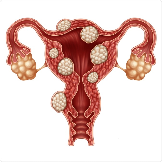
UTERINE FIBROIDS
By A.A. (staff writer) , published on October 25, 2021
Medicine Telehealth Health fibroids uterine fibroids


The noncancerous growths in the uterus that usually occur during pregnancy are called uterine fibroids. They are also known as leiomyomas or myomas, aren't linked to an elevated risk of uterine cancer, and usually never turn cancerous. Fibroids may differ in size from tiny seedlings that are undetectable to enormous masses that deform and expand the uterus. A single fibroid or a group of them can be present. Multiple fibroids can cause the uterus to enlarge to the point where it reaches the rib cage, causing weight gain.
Uterine fibroids affect many women at some point in their life. However, because uterine fibroids rarely show symptoms, you may be unaware that you have them. During a pelvic exam or a pregnancy ultrasound, your doctor may find fibroids by chance.
Symptoms
Symptoms might not be present in the majority of women who have fibroids. On the other hand, if symptoms are present then they depend on the number of fibroids, size, and location. The following are the most common signs and symptoms experienced by women having uterine fibroids.
- Extended menstrual periods that last more than a week.
- Heavy menstrual bleeding.
- Pain or pelvic pressure
- Frequent urination
- Leg pains or backache
- Problem with emptying the bladder
- Rarely, a fibroid can cause acute pain when it outgrows its blood supply and begins to die.
Mostly, fibroids are identified by their location. Fibroids that grow within the muscular uterine wall are referred to as intramural fibroids. The uterine cavity is engulfed by submucosal fibroids. Subserosal fibroids protrude beyond the uterus's walls1.
Diagnosis of fibroids
You must consult a gynecologist for a pelvic exam to acquire an accurate diagnosis. The purpose of this inspection is to determine the health, size, and form of your uterus. You may also require additional tests, such as:
Ultrasound
High-frequency sound waves are used to create pictures of your uterus on a screen during an ultrasound. It will enable your physician to examine the interior components of the tumor as well as any fibroids that may be present. Because the ultrasound wand is placed into the vagina, a transvaginal ultrasound may offer sharper images because it is closer to the uterus.
Pelvic MRI
This in-depth imaging test creates pictures of the ovaries, uterus, and pelvic organs2.
Computed tomography (CT):
A CT scan makes a comprehensive image of your inside organs from many angles using X-ray imaging.
Hysteroscopy:
Your doctor will use a tool which is a thin, flexible tube with a camera on the end known as a scope to check for fibroids inside your uterus during a hysteroscopy. The scope is inserted into your uterus after passing through your vaginal and cervix.
Hysterosalpingography (HSG):
This is a comprehensive X-ray in which contrast material is injected first, followed by X-rays of the uterus. It is more commonly used in women who are simultaneously being evaluated for infertility.
Sonohysterography:
A tiny catheter is inserted transvaginally, and saline is administered into the uterine cavity via the catheter. This additional fluid aids in the creation of a sharper image of your uterus as compared to conventional ultrasound.
Laparoscopy:
Your doctor will create a tiny incision in your lower abdomen throughout this test. A small, flexible tube containing a camera on the end will be implanted to examine your inside organs in more detail3.
Types of fibroids
Treatment suggestions are influenced by the kind of fibroids as well as their size and quantity. Fibroids are divided into three categories:
Subserosal fibroids:
This kind of fibroids is the most prevalent. They have the ability to push through the uterus and into the pelvis. Subserosal fibroids can increase in size and have a stalk that connects to the uterus at times (pedunculated fibroid).
Intramural fibroids:
Intramural fibroids are formed in the muscular wall of the uterus.
Submucosal fibroids:
Submucosal fibroids are rare. They can develop into the uterus's open region and may even have a stalk.
Causes of fibroids
Uterine fibroids have some unknown causes. Moreover, some research studies show that it is linked with genetic components. There is no specific external stimulation that triggers a woman to grow fibroids.
Risk for uterine fibroids
There are many risk factors associated with the development of uterine fibroids
Age:
Fibroids are more prevalent in females as they become older, specifically in their 30s and 40s and till menopause. Fibroids are significantly less likely to form after menopause, and if they do, they normally diminish with the passage of time.
Family history:
The chances of fibroids increases when a family member has a history of fibroids. The risk of getting uterine fibroids ate higher if the woman's mother had a history of fibroids.
Ethnic origin:
The risk of fibroids is higher in African-American women than in other ethnicities.
Obesity:
Obese women are more likely to have fibroids. The danger is two to three times higher for very overweight women than for typical women4.
References
- Uterine fibroids. [Internet]. [Cited 2021 July 12]; Available from https://www.mayoclinic.org/diseases-conditions/uterine-fibroids/symptoms-causes/syc-20354288
- Fibroids. [Internet]. [Cited 2021 July 12]; Available from https://www.healthline.com/health/uterine-fibroids#treatment
- Uterine fibroids. [Internet]. [Cited 2021 July 12]; Available from https://my.clevelandclinic.org/health/diseases/9130-uterine-fibroids
- What are fibroids? [Internet]. [Cited 2021 July 12]; Available from https://www.uclahealth.org/fibroids/what-are-fibroids
Find articles related to: Medicine Telehealth Health fibroids uterine fibroids
More articles about Female Topics
Back to the Health Tips Index